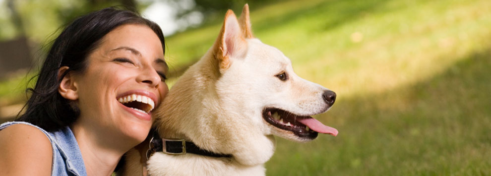
Cranial Cruciate Ligament
Cranial Cruciate Ligament (CrCL) disease is a very common cause of hindlimb lameness in dogs, especially medium to large breeds. The CrCL is the equivalent to the ACL (anterior cruciate ligament) in humans and connects the femur (thigh bone) to the tibia (shin bone) so stabilising the knee joint and preventing rotation or over extension. It is a very tough band of tissue. In humans, the ligament usually ruptures due to a sudden trauma but in most dogs the CrCL ruptures as a result of long-term degeneration with the ligament fibres weakening over time. In dogs there is a strong genetic cause with some animals rupturing both ligaments at a relatively early age. Individual conformation, obesity and hormonal changes could also play a role.
The most common symptom is a hindlimb lameness. This can be sudden onset at exercise (e.g. chasing a fox down the garden) or more intermittent and progressive. Dogs will often struggle to get up after resting especially if the condition is bilateral. The fraying ligament means there is joint instability so osteoarthritis develops rapidly due to abnormal wear within the joint. This instability is not only painful but causes a mechanical lameness due to the increased movement within the joint and the femur slipping backwards against the tibia. This movement can also tear the menisci within the knee that normally act as a shock absorber. The most common meniscus to be injured is the medial one on the inside of the joint.
A diagnosis of a full rupture of the CrCL is made by the vet examining the knee joint for laxity. The vet will assess the stifle for a cranial drawer which means the femur can be moved relative to the tibia. This is often not easy to perform on a conscious dog as they may be in a lot of pain and tense the surrounding muscles, so it can be carried out under sedation in combination with an X-ray to assess other changes within the knee joint. Partial tears of the ligament are not always easy to diagnose, they may need to be confirmed with an MRI scan or arthroscopy (a putting a camera into a joint).
Treatment can be surgical or medical. Non-surgical options have limitations and are associated with poorer long-term outcomes. Generally, medical options are only used for smaller dogs, if there is a significant risk with a general anaesthetic (e.g. a heart condition or immune disorder) or, if the owner has financial constraints. Medical options are more commonly used for partial rather than complete tears and involves weight management, physiotherapy, long-term anti-inflammatory painkillers and exercise modification. Dogs under 15kgs can regain a good quality of life with medical management but rarely attain complete recovery.
Dogs over 15kg have a poor chance of recovery without surgery. There are 2 varying surgical techniques; one aims to replace the torn ligament, and the other involves cutting the tibia and altering the forces on the stifle joint. There are numerous ligament replacement techniques. Some use the transfer of local fascia, but this technique is only used in smaller dogs as it is prone to failure as the replacement tissue is not as strong as the original ligament and is in the same unfavourable position that predisposed the ligament failing in the first place. They also tend to limit range of movement. Prosthetic ligament replacement techniques are more successful and enable a more normal return to limb movement but are often not robust enough in heavy or athletic dogs. They are most useful in purely traumatic ligament injuries where there is little or no anatomical abnormality.
The most successful surgeries alter the angles of the knee joint by cutting the tibia and fixing it in a new position to enable stability in the knee joint without a CrCL . The most common surgery is th